AP SHOULDER
Anteroposterior shoulder projection protocol
Exposure Factors
Medium-low exposure: Parameters for optimal visualization of shoulder joint
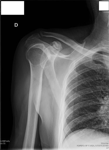
Anatomical Structures Visible
Should be clearly observed:
- Shoulder joint (glenohumeral)
- Complete clavicle
- Proximal humerus (head and neck)
- Part of scapula (acromion, scapular spine)
- Glenohumeral joint space
Field verification:
Proper field: Include proximal humerus and complete clavicle in same image
Cassette Size and Orientation
Transverse orientation to cover shoulder joint and adjacent structures
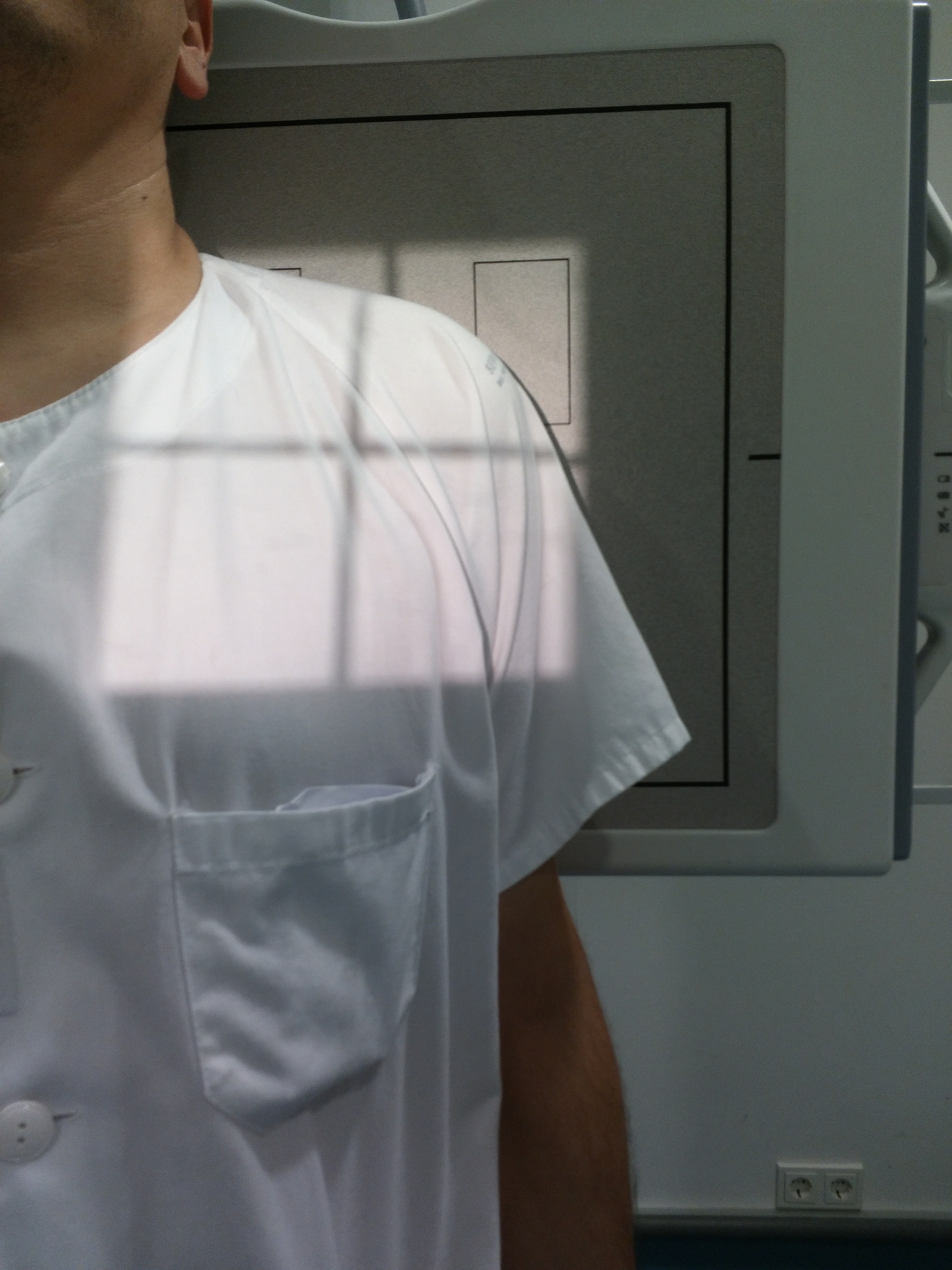
Patient Positioning
Central Ray Point
Direction: Horizontal and perpendicular to shoulder joint
Location: At level of coracoid process of scapula, centered on cassette
Objective: Optimal glenohumeral joint visualization
Specific Hand Position
Correct Position
Hand at side of body
Palm of hand against thigh
Arm in neutral rotation
Epicondylar Alignment
Line between epicondyles at 45° over cassette plane
Avoids structure superposition
Improves joint visualization
Patient Instructions
"Do not breathe during the exposure"
Maintain position without movement and apnea during radiographic exposure
1. "Turn your body slightly toward the affected side"
2. "Keep your hand resting against your thigh"
3. "Stay completely still"
4. "Take a deep breath and hold it"
5. "Do not move during the exposure"
Optimal Image Characteristics
Joint Space
Glenohumeral clearly visible
No Superposition
Differentiated bone structures
Alignment
Humeral head centered in glenoid
Full Field
Proximal humerus to clavicle
Common Technical Challenges
Frequent problems in AP shoulder projection:
- Structure superposition from incorrect arm rotation
- Insufficient body rotation not placing shoulder flat against bucky
- Incorrect hand position affecting humeral rotation
- Exclusion of structures from poor cassette placement
- Patient movement during exposure
Solution: Verify adequate body rotation and hand position with palm against thigh
Special Considerations
Painful Patient
Position with maximum possible comfort, adjust rotation according to tolerance.
Geriatric Patient
Consider mobility limitations, possible need for additional support.
Pediatric Patient
Reduce exposure according to age and ALARA protocol, adjust cassette size.